Toby presented to his regular veterinarian after an altercation with a pig. There was a 3 cm laceration to the side of the face over the zygomatic arch. The wound healed with open wound management and a short course of antibiotics, however Toby represented with a fluid filled mass on the side of his face caudal to the initial injury 1 month later. The referring veterinarian aspirated the mass and found fluid consistent with saliva and he was referred for further investigation.
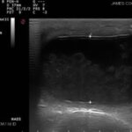
Ultrasound
An ultrasound guided aspirate of the mass revealed a thick mucoid yellow tinged fluid. Cytology of the fluid revealed eosinophilic stands, occasional mature neutrophils and sparse epithelial cells.
Sialography
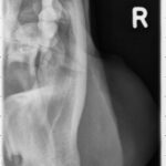
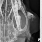
Sialography by catheterisation of the parotid duct confirmed a sialocele. Figure 2a demonstrates the plain radiographs, Figure 2b demonstrate the sialogram and Figure 2c demonstrates the placement of the catheter. Recognised retrospectively was the foreign body cranial to the sialocoele.
Surgical Procedure
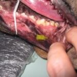
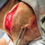
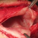
A small 2cm incision was made over the fibrous lump rostral to the sialocoel. Blunt dissection into this area revealed a small 1cm length piece of bony material consistent with a pig tusk that was subsequently removed and the sialocele drained (Figure 3). Removal of the parotid gland was performed preserving the facial nerve.
Conclusion
Toby made an uneventful recovery without recurrence of the sialocele.
Submitted by: Dr. Craig Thomson (surgical resident)

