Type I IVDD is a common canine spinal disease that can affect a large variety of breeds but is more common in young to middle-aged chondrodystrophic breeds. In Part 1 we discussed neurological examination, neurolocalisation and grading. In Part 2 of this blog we will review the diagnosis, management and prognosis of type I IVDD.
Differentials
Canine spinal disease or IVDD is one of the more common causes for hindlimb ataxia but this does not mean other differentials should not be considered. It can be helpful to categorise the differentials to ensure that none are missed. The DAMNIT-V scheme is probably the most commonly used.
| Differential for T3-L3 spinal lesion | |
| Degenerative | IVDD, degenerative myelopathy, synovial cysts, bilateral cruciate disease |
| Anomalous | Spinal arachnoid cyst, vertebral malformation (including hemivertebrae) |
| Metabolic | Myasthenia gravis, Addison’s disease, hypoglycaemia |
| Neoplastic/Nutritional | Primary or secondary tumours, hypervitaminosis A, thiamine deficiency |
| Idiopathic/Infectious/Iatrogenic | DISH, discospondylitis, GME/MUO/other meningitides, foreign body migration, spinal epidural empyema, surgical injury |
| Traumatic/Toxic | Traumatic disc injury, fracture/luxation, toxin ingestion |
| Vascular | FCE, neurogenic claudication, spinal cord haemorrhage |
This is not an exhaustive list but a list of the more common differentials. The bold type in the tables are the more common differentials that we see.
Diagnostic work up
As shown in the list above the differentials can be long but the signalment, history and physical examination should rule in or out a lot of these. A classic type I IVDD occurs as an acute onset clinical sign in young to middle-aged chondrodystrophic dogs. Plain film radiographs are useful in ruling out discospondylitis, obvious neoplasia as well as vertebral fractures or luxations. Radiographic evidence of spondylosis deformans does not appear to be associated with extruded discs. It does not allow for accurate identification of compressive disc disease. Myelographic accuracy is reported between 50-98% but remember the complications associated with it (seizures 10-22%, apnea, cardiac arrhythmias, meningitis, subarachnoid haemorrhage).
The next question to answer is whether to proceed with advanced imaging MRI or CT. Gold standard advance imaging would be an MRI. In most cases, a CT gives us the information we need, in a recent study it was shown to show IVDD in over 90% of Daschunds and over 80-85% of other small breeds. CT is better for imaging bone or mineralised material i.e. extruded or protruded IVDD. CT is faster, more readily available as well as cheaper compared to an MRI. MRI will show soft tissue detail, i.e. details of the spinal cord odema/haemorrhage and the degree of compression. If disease other than a classic extruded acute IVDD we would recommend an MRI.

Treatment
The treatment options for canine spinal disease are conservative management vs surgery. The decision as to which one will be performed is based mostly on the findings of the neurological examination and advanced imaging, but the client’s decision both for rehabilitation and finances need to be taken into consideration. Surgery is generally indicated if the pain is not controlled with conservative management or if the animal is non-ambulatory. Surgery will also allow for a faster recovery.
Conservative management consists of strict exercise restrictions with regular physiotherapy and analgesia. The aim of this is to allow for the secondary changes occurring within the injured spinal cord to subside as well as the material within the canal to settle and fibrose to prevent further compression of the spinal cord. For analgesia we commonly combine NSAIDS, Gabapentin as well as opioids and Acetaminophen as needed.
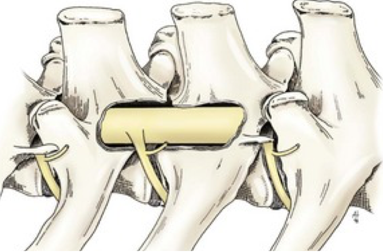
Surgical management normally involves decompression with a hemilaminectomy at the site of the disc extrusion. A hemilaminectomy can be performed through a dorsal or a dorsolateral approach over the spinous processes of the site of protrusion and generally 1-2 processes caudally and cranially for exposure. The lateral vertebrae are exposed and the hemilaminectomy window created using a high-speed burr. The extruded disc material is removed with care and gentle manipulation using fine spinal instruments until the spinal cord has resumed its normal position. Sometimes we would perform disc fenestration and/or durotomy for further decompression. Postoperative management is similar to conservative – strict exercise restriction for 4-6wks with physiotherapy and analgesia.

Prognosis
The prognosis of thoracolumbar disc extrusion in dogs and return to ambulation varies greatly depending on the grade/severity of clinical signs at presentation. The table below gives an indication of the mean proportion of dogs that recover based on the grading as well as treatment type from a recent systemic review paper (Langerhuus L et al Vet J 2017)
| Neurological Grade | Conservative Management | Surgical Management |
| 3 | 79% | 93% |
| 4 | 62% | 93% |
| 5 | 10% | 61% |
Canine spinal disease can be difficult as diagnosis requires advanced imaging which is not always readily accessible to primary care vets. This blog hopefully will fill in some gaps to allow a more confident diagnosis of thoracolumbar spinal extrusion and to be able to give clients an idea of prognosis with both conservative and surgical management of this condition.
Remember there are other spinal diseases that we did not discuss here including type II intervertebral disc protrusion (occurring more in older non-chondrodystrophic dogs), acute non-compressive nucleus pulposus extrusion, hydrated nucleus pulposus extrusion, extradural synovial cysts which can all present similarly.
If you have any questions or would like to view any of the articles or figures from within this blog feel free to contact us at the clinic and we would be happy to send them through.

