Leo the Cat
by Dr Tony Caiafa (Veterinary Practitioner in Dentistry)
This case study emphasises the importance of a complete oral health assessment and treatment (COHAT) combined with intraoral radiography when diagnosing and treating oral diseases in companion animals.
“Leo” is a 12-years old neutered male domestic long hair cat presented with oral discomfort and a chronic skin problem at Northside Vet Care. He had previously been treated at the same practice for an oral problem three months earlier. At that time, treatment consisted of crown amputation of left maxillary canine tooth (#204) due to a suspected type 2 tooth resorption (TR) and a dental clean.
Stage 1: Conscious Examination of “Leo”:
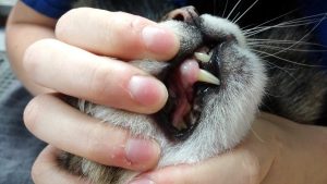
A detailed history was taken from the owner and an awake examination was performed. Leo was mildly resentful of inspection of the oral cavity which may have indicated that oral discomfort/pain was still present. Regardless of the recent treatment history of dental clean and crown amputation, overall dental disease score was PD1 (based on this awake examination). There was a complicated crown fracture of tooth #404 (pulp exposure) and a possible pulp exposure involving the fractured crown of tooth #304. There was an uncomplicated crown fracture involving #104 also.
Picture 1: Complicated crown fracture on #404. Uncomplicated crown fracture of #104 is also shown.
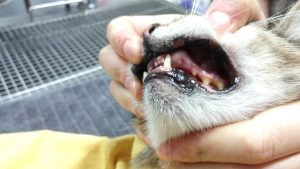
Picture 2: Showing ulceration of edge of upper left lip (blue arrow) as well as complicated crown fracture of #304. In this picture also shows absence of #101.The ulceration on the upper lip was due to lip entrapment from tooth #304
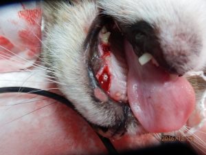
A small ulcer involving the left upper lip (size approx. 4mm in diameter) was noted, which may have been caused by trauma from the fractured left mandibular canine tooth (#304). The #204 site had not completely healed and there was still gingival ulceration present here. The left maxillary canine tooth (#204) previously had a crown amputation procedure (see picture). The treatment was based on a diagnosis of type 2 resorption (possible loss of periodontal ligament space on an intraoral radiograph of this tooth). He was showing signs of pain when the area was touched with a cotton Q tip. Mild swelling of the gingiva in the area of #204/#206 was also noted during the examination.
The owner was also concerned about Leo’s thinning of the coat around his hind limbs. Small, multiple scabs around the head were observed during the conscious examination. Otherwise, all other physical examination including head and neck was normal as well as all vitals were within normal limit during the physical examination. Considering the age of Leo, and although blood tests had been done three months earlier, pre-anaesthetic laboratory tests, including complete blood count (CBC) and biochemistry, including total T4 and urine specific gravity were repeated. The high end of normal creatinine level was observed. However, all other laboratory findings were within normal range.
Treatment Planning:
Due to multiple abnormal findings noted in Leo’s mouth, comprehensive examination and treatments under the general anaesthesia (COHAT) were strongly recommended. The owner wished to preserve Leo’s teeth if possible. Due to this wish by the owner, root canal treatments (RCT) were offered for the complicated crown fractures teeth #304/404. A intraoral radiographic assessment was imperative to assess the pathology involving these teeth.
Stage 2: Oral Examination under General Anaesthesia
“Leo” was given general anaesthesia for extra and intraoral examination. Detailed, tooth-by-tooth examination by using periodontal probe and explorer tip revealed multiple complex problems in the oral cavity. Multiple fleas were also found on the coat during examination and treatment under general anaesthetic. Due to the complexity and extensive nature of the dental disease see in the patient, whole mouth intraoral dental radiographs were performed using the latest available technologies (Portex hand held Xray generator, iM3 Sydney, CR7 digital dental Xray processor, iM3 Sydney), and treatment plan was established based on the examination finding.
Stage 3: Diagnosis Oral Disease
Diagnosis and treatments of Leo’s dental disease as follows:
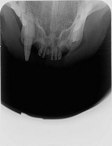
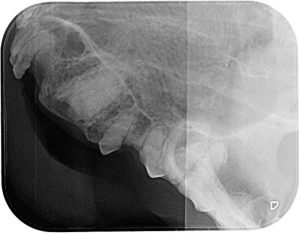
Tooth number #101 showed an apical lucency associated with the retained root
Picture 3: fractured crown retained root #101 with apical lucency
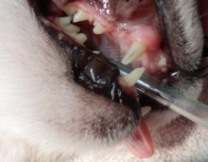
Tooth resorption (TR) was diagnosed by probing of #107. Intraoral dental radiography shows type 1 TR. Complete extraction was indicated for the treatment of TR.
Picture 4: Clinical pictures showing uncomplicated crown fracture of #107
Picture 5: showing TR type 1 – Note clear PDL of mesial root. Zygomatic arch is overlapping on distal root of #107.
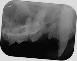
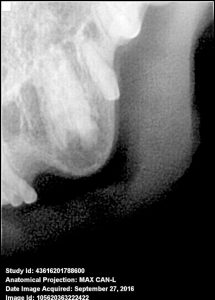
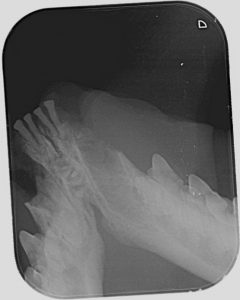
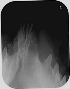
Tooth #204 was re-examined by repeating radiographic examination. It revealed Type 1 Tooth Resorption rather type 2, with an intact periodontal ligament space around this tooth root, thus complete root extraction was indicated.
Picture 6 (Left): Lateral view intraoral radiograph (slightly elongated) of crown amputated tooth #204 (DR senro, EVA Vet™, USA).
Picture 7 (right): same tooth as in picture 6, but lateral view intraoral radiograph taken using CR phosphor plate size 2 (CR7 X-ray Processor, iM3, Sydney)
Note the differences in image quality between the two images taken using the same X-ray generator (Portex hand held X-ray generator, iM3, Sydney). The image contrast and image quality are better in picture 7, showing the periodontal ligament space more clearly. This would suggest that tooth resorption involving #204 is a type 1 TR (complete extraction required, not crown amputation) and not a type 2.There is also under and overexposure of the image in picture 6 which can be seen when using DR sensors. This can sometimes mask pathology.
Tooth #304 appears to have an external resorption, possibly due to tooth resorption or inflammatory root resorption associated with a necrotic pulp which is not a good candidate to perform RCT. Thus, extraction was indicated.
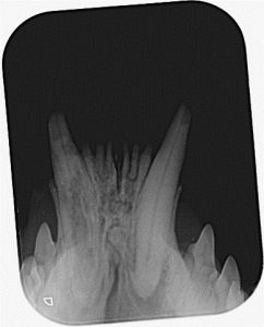
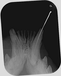
Picture 8A (left) and 8B (right): Showing inflammatory root resorption of tooth #304. It is most likely associated with pulp necrosis. Picture 8B: Shows an endodontic file unable to negotiate the root canal system of tooth #304. It is most likely associated with pulp necrosis. Picture 8B shows an endodontic file unable to negotiate the root canal system of tooth #304, suggestive of internal combined with external inflammatory resorption. The entire tooth would need extracting based on these intraoral images
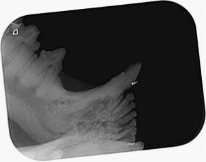
Tooth #404 shows apical pathology due to pulp necrosis. Extraction was indicated.
Picture 9A (left) and 9B (right): showing complicated crown fracture and apical pathology associated with tooth #404 (canine tooth on right side of image). Extraction was indicated due to the long standing nature of the pulp necrosis and failure to negotiate the whole of the root canal system with a file, suggesting apical sclerosis of the canal (picture 9B)
Leo’s treatment:
All teeth/roots extractions required labial/buccal mucoperiosteal flaps and removal of labial/buccal and, in some instances, lingual bone. All extraction sockets were curetted and flushed with saline prior to suturing. This was to make certain that no granulation tissue of bony debris was left behind in the socket that may lead to a non- healing extraction site. All flaps were sutured with 5/0 Vicryl Rapide (Polyglactin 910, multi-filament, absorbable sutures: Ethicon). All extraction sockets were radiographed to confirm complete removal of the extracted teeth.
The patient was given perioperative dose of amoxicillin/clavulanic acid antibiotic and local anaesthetic nerve blocks (caudal maxillary and caudal mandibular nerve blocks with 2% lignocaine HCl at dose of 2mg/Kg). Meloxicam was given subcutaneously as post-operative analgesia (0.2mg/kg).
The patient was sent home with 7 days course of oral amoxicillin/clavulanic acid (75mg), meloxicam pain relief for 48 hours as well as instructed to treat fleas by using Revolution.
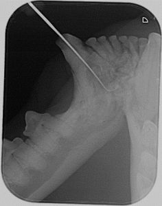
Picture 10: showing surgical exposure (envelope flap) of the root of tooth #204. A gutter was created with a surgical length fissure bur in a LED high speed handpiece (iM3, Sydney). This gutter allowed for the introduction of small winged dental elevators (winged elevators, iM3, Sydney) and extraction of the entire root.
Picture 11A (left) and 11B (right): Perioperative radiograph of teeth #304 and #404 showing complete removal of #404 and root remaining in #304. A gutter was created around the remaining root of #304 and the root elevated out (picture 11B)
Diet was strictly soft food only for 2 weeks and advised minimal handling of mouth for 2 weeks.
Post-operative follow ups over a 6 week period, showed that Leo was a much happier cat without any mouth pain. All surgical extraction sites healed uneventfully.
Conclusions:
This case highlights the importance of detailed and complete oral examination including whole mouth intraoral dental radiographs in an elderly cat. Good quality intraoral dental radiographs are vital to assess the types of tooth resorption which will dictate the type of treatment instigated.
Intraoral radiography is one of the most effective diagnostic tools for the investigation of missing tooth/teeth, fractured teeth as well as treatment planning for tooth resorptions to determine when crown amputation or full extraction is appropriate.

